Hsv Encephalitis Mri Findings - Contribution Of Mri In Serious Forms Of Acute : Herpes simplex (hsv) encephalitis is the most common cause of fatal sporadic fulminant necrotising viral encephalitis and has characteristic imaging findings.
Get link
Facebook
X
Pinterest
Email
Other Apps
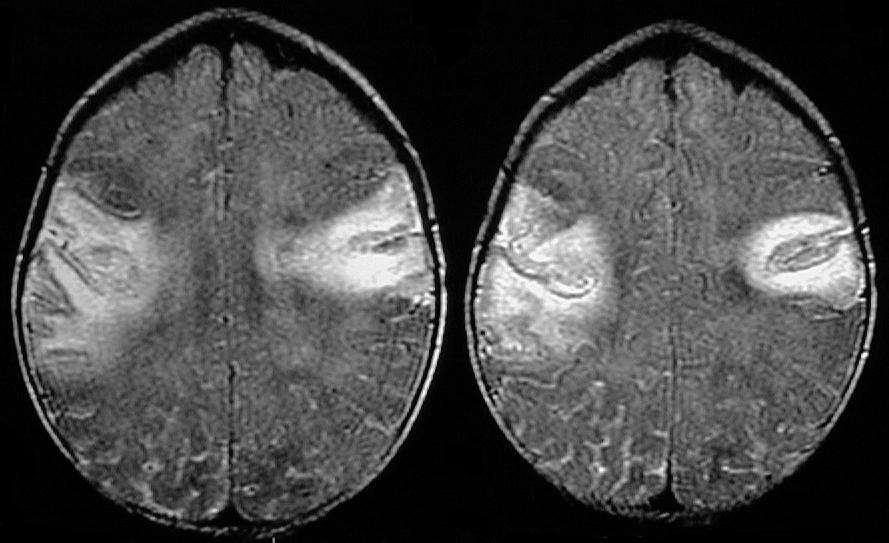
Hsv Encephalitis Mri Findings - Contribution Of Mri In Serious Forms Of Acute : Herpes simplex (hsv) encephalitis is the most common cause of fatal sporadic fulminant necrotising viral encephalitis and has characteristic imaging findings.. Normal mr imaging findings (type 1, 53% of patients), isolated hippocampal involvement (type 2, 13%), other brain lesions without hippocampal involvement (type 3, 13%), and other brain lesions with hippocampal involvement (type 4, 21%). Lesions similar to cytotoxic edema, and lesions similar to vasogenic edema. Neonatal herpes simplex encephalitis is caused by vertical transmission of infection during passage from birth canal with diffuse cerebral involvement within the first month after birth; The patients with the former type of lesions had fulminating disease, and were in severe clinical condition. In patients with herpes encephalitis, two distinct types of diffusion imaging findings (on b =1000 s/mm 2 images, and adc maps) were noted:
The clinical syndrome is often characterized by the rapid onset of fever, headache, seizures, focal neurologic signs, and impaired consciousness 1 . Two subtypes are recognised which differ in demographics, virus, and pattern of involvement. Hatipoglu hg(1), sakman b, yuksel e. On pathology, herpes viruses cause a fulminant hemorrhagic and necrotizing meningoencephalitis, with typical gross findings of severe edema and massive tissue necrosis, with petechial hemorrhages. The patients with the former type of lesions had fulminating disease, and were in severe clinical condition.
An Update On Diagnostic Imaging Studies For Viral Encephalitis Future Virology from www.futuremedicine.com Watershed distribution ischemia in areas remote from the primary herpetic lesions may be seen. Normal mr imaging findings (type 1, 53% of patients), isolated hippocampal involvement (type 2, 13%), other brain lesions without hippocampal involvement (type 3, 13%), and other brain lesions with hippocampal involvement (type 4, 21%). On pathology, herpes viruses cause a fulminant hemorrhagic and necrotizing meningoencephalitis, with typical gross findings of severe edema and massive tissue necrosis, with petechial hemorrhages. For viral infection of the meninges, please refer to the general article on viral meningitis, and, for a broad view on the curriculum of infections of the central nervous system, refer to cns. Therefore, recognition of the mr imaging. In their important analysis, the authors describe 4 distinct imaging patterns: Lesions similar to cytotoxic edema, and lesions similar to vasogenic edema. Hatipoglu hg(1), sakman b, yuksel e.
Lesions similar to cytotoxic edema, and lesions similar to vasogenic edema.
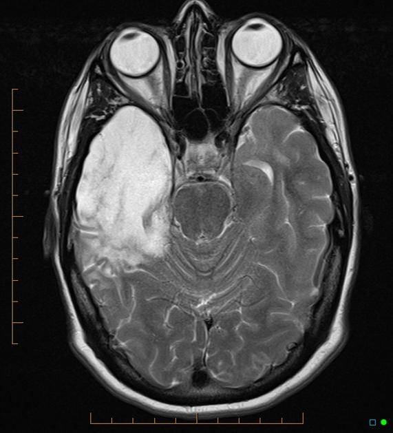
Lesions similar to cytotoxic edema, and lesions similar to vasogenic edema. Seizure, altered sensorium, fever frontal and temporal lobes, rarely extratemporal t2 hyperintensity, restricted diffusion, sometimes. Herpes encephalitis is the most common sporadic encephalitis in the united states and other industrialized countries .occurring as either a primary infection with herpes simplex virus or as a reactivation of latent virus, herpes encephalitis causes significant morbidity and mortality .early intervention with acyclovir significantly improves outcome; The clinical syndrome is often characterized by the rapid onset of fever, headache, seizures, focal neurologic signs, and impaired consciousness 1 . Having said that, mri with contrast is considered the most sensitive imaging modality, and findings are present in over half of individuals 8. The patients with the former type of lesions had fulminating disease, and were in severe clinical condition. Herpes simplex encephalitis occurs as 2 distinct entities: 4 it was found that, rather surprisingly, varicella zoster virus (vzv), the cause of chickenpox and herpes zoster, was the most frequently detected virus at 29%, with hsv and enteroviruses accounting for 11% of cases. Two subtypes are recognized which differ in demographics, virus, and pattern of involvement. (1)department of radiology, ankara numune education and research hospital, ankara, turkey. Neonatal herpes simplex encephalitis is caused by vertical transmission of infection during passage from birth canal with diffuse cerebral involvement within the first month after birth; As the older term limbic encephalitis implies, the most common location of involvement is the mesial temporal lobes and limbic systems, typically manifested by cortical thickening and increased t2/flair. In patients with herpes encephalitis, two distinct types of diffusion imaging findings (on b =1000 s/mm 2 images, and adc maps) were noted:
mri findings of herpes simplex encephalitis. Lesions similar to cytotoxic edema, and lesions similar to vasogenic edema. Normal mr imaging findings (type 1, 53% of patients), isolated hippocampal involvement (type 2, 13%), other brain lesions without hippocampal involvement (type 3, 13%), and other brain lesions with hippocampal involvement (type 4, 21%). Comparison with findings of herpes simplex virus encephalitis. We compared the imaging findings of the two conditions.
Radiology In Ped Emerg Med Vol 7 Case 9 from www.hawaii.edu For viral infection of the meninges, please refer to the general article on viral meningitis, and, for a broad view on the curriculum of infections of the central nervous system, refer to cns. Two subtypes are recognized which differ in demographics, virus, and pattern of involvement. A recent study in finland also used pcr to detect various viruses in the csf of over 3000 patients who had infections of the cns including encephalitis, meningitis, and myelitis. The patients with the former type of lesions had fulminating disease, and were in severe clinical condition. Despite advances in antiviral therapy over the past 2 decades, herpes simplex encephalitis (hse) remains a serious illness with significant risks of morbidity and death. Normal mr imaging findings (type 1, 53% of patients), isolated hippocampal involvement (type 2, 13%), other brain lesions without hippocampal involvement (type 3, 13%), and other brain lesions with hippocampal involvement (type 4, 21%). Lesions similar to cytotoxic edema, and lesions similar to vasogenic edema. As the older term limbic encephalitis implies, the most common location of involvement is the mesial temporal lobes and limbic systems, typically manifested by cortical thickening and increased t2/flair.
Herpes simplex (hsv) encephalitis is the most common cause of fatal sporadic fulminant necrotising viral encephalitis and has characteristic imaging findings.
We present mri findings of two cases of herpes simplex encephalitis (hse) confirmed by pcr analysis, focusing on the serial changes after acyclovir therapy: 4 it was found that, rather surprisingly, varicella zoster virus (vzv), the cause of chickenpox and herpes zoster, was the most frequently detected virus at 29%, with hsv and enteroviruses accounting for 11% of cases. mri findings of herpes simplex encephalitis. For viral infection of the meninges, please refer to the general article on viral meningitis, and, for a broad view on the curriculum of infections of the central nervous system, refer to cns. The patients with the former type of lesions had fulminating disease, and were in severe clinical condition. The kappa value for interobserver agreement on rating the scans as normal or abnormal was good (0.65) for ct and moderate (0.59) for mri. Noguchi t, yoshiura t, hiwatashi a, et al. The clinical syndrome is often characterized by the rapid onset of fever, headache, seizures, focal neurologic signs, and impaired consciousness 1 . The patients with the former type of lesions had fulminating disease, and were in severe clinical condition. Specific diagnosis often requires pcr. As the older term limbic encephalitis implies, the most common location of involvement is the mesial temporal lobes and limbic systems, typically manifested by cortical thickening and increased t2/flair. In patients with herpes encephalitis, two distinct types of diffusion imaging findings (on b =1000 s/mm 2 images, and adc maps) were noted: A recent study in finland also used pcr to detect various viruses in the csf of over 3000 patients who had infections of the cns including encephalitis, meningitis, and myelitis.
Specific diagnosis often requires pcr. Normal mr imaging findings (type 1, 53% of patients), isolated hippocampal involvement (type 2, 13%), other brain lesions without hippocampal involvement (type 3, 13%), and other brain lesions with hippocampal involvement (type 4, 21%). In patients with herpes encephalitis, two distinct types of diffusion imaging findings (on b =1000 s/mm 2 images, and adc maps) were noted: Watershed distribution ischemia in areas remote from the primary herpetic lesions may be seen. article in japanese yoshioka a, hirose g, tsukada k, oda r, kosoegawa h.
Temporal Lobe Atrophy Post Herpes Simplex Encephalitis Radiology Case Radiopaedia Org from prod-images-static.radiopaedia.org (1)department of radiology, ankara numune education and research hospital, ankara, turkey. article in japanese yoshioka a, hirose g, tsukada k, oda r, kosoegawa h. Two subtypes are recognised which differ in demographics, virus, and pattern of involvement. Seizure, altered sensorium, fever frontal and temporal lobes, rarely extratemporal t2 hyperintensity, restricted diffusion, sometimes. Encephalopathy from herpes simplex encephalitis (hse). Specific diagnosis often requires pcr. Having said that, mri with contrast is considered the most sensitive imaging modality, and findings are present in over half of individuals 8. We present mri findings of two cases of herpes simplex encephalitis (hse) confirmed by pcr analysis, focusing on the serial changes after acyclovir therapy:
Therefore, recognition of the mr imaging.
The patients with the former type of lesions had fulminating disease, and were in severe clinical condition. The diagnosis of hse is based on laboratory investigations, magnetic resonance (mr) imaging findings, electroencephalogram and, occasionally, a biopsy. Herpes encephalitis is the most common sporadic encephalitis in the united states and other industrialized countries .occurring as either a primary infection with herpes simplex virus or as a reactivation of latent virus, herpes encephalitis causes significant morbidity and mortality .early intervention with acyclovir significantly improves outcome; Herpes simplex (hsv) encephalitis is the most common cause of fatal sporadic fulminant necrotising viral encephalitis and has characteristic imaging findings. In patients with herpes encephalitis, two distinct types of diffusion imaging findings (on b =1000 s/mm 2 images, and adc maps) were noted: Relapse of encephalitis occurs in up to 10% of patients, manifested by recurrent symptoms, clinical and mri findings, and the presence of herpes simplex virus type 1 dna in the cerebrospinal fluid (csf). In children older than 3 months and in adults, hse is usually localized to the temporal and frontal lobes and is c. In patients with herpes encephalitis, two distinct types of diffusion imaging findings (on b =1000 s/mm 2 images, and adc maps) were noted: Herpes simplex (hsv) encephalitis is the most common cause of fatal sporadic fulminant necrotizing viral encephalitis and has characteristic imaging findings. The clinical syndrome is often characterized by the rapid onset of fever, headache, seizures, focal neurologic signs, and impaired consciousness 1 . We present mri findings of two cases of herpes simplex encephalitis (hse) confirmed by pcr analysis, focusing on the serial changes after acyclovir therapy: Two subtypes are recognised which differ in demographics, virus, and pattern of involvement. Despite advances in antiviral therapy over the past 2 decades, herpes simplex encephalitis (hse) remains a serious illness with significant risks of morbidity and death.
Herpes simplex (hsv) encephalitis is the most common cause of fatal sporadic fulminant necrotizing viral encephalitis and has characteristic imaging findings hsv encephalitis mri. article in japanese yoshioka a, hirose g, tsukada k, oda r, kosoegawa h.

Comments
Post a Comment